1
/
of
1
Oxford University Press, USA
Brain Imaging: A Guide for Clinicians
Brain Imaging: A Guide for Clinicians
Regular price
$145.00 USD
Regular price
Sale price
$145.00 USD
Shipping calculated at checkout.
Quantity
Couldn't load pickup availability
Brain Imaging: A Guide for Clinicians is designed to provide a foundation of information necessary for those wishing to integrate brain imaging into their practice, or to those who currently review brain scans but have minimal formal training in neuroimaging. The guide covers a range of topics
important to those using brain imaging, such as the strengths and weaknesses of the many different techniques currently available, the factors that may influence the use of imaging data, common pitfalls or artifacts that may be misleading to the clinician, the most appropriate techniques to use
given a specific clinical question or condition, how to interpret information presented on a brain image, and also how many pathological conditions appear on a variety of brain scanning techniques or sequences. This guide also provides detailed information regarding the identification of primary
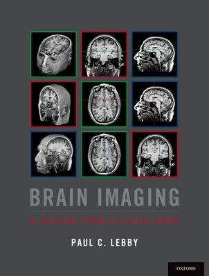
brain regions, anatomical structures, systems or pathways using both two-dimensional and three-dimensional imaging techniques. A brain atlas is included using both CT and MRI sequences to facilitate the reader's ability to identify most primary brain structures. A novel color-coded system is used
throughout this guide to assist the reader in identifying slice locations and orientations. Images with green borders are displayed in the axial plane, with the slice location being shown on other orthogonal image planes by a green line. Similarly, images with a red border are displayed in the
coronal plane and those with a blue border are displayed using a sagittal plane; red and blue reference lines are displayed on orthogonal slices to identify the slice location. The crosshairs formed by the color-coded reference lines optimize the reader's ability to identify primary anatomical
structures or pathological markers and processes. Chapters in this book progress from a general description of the clinical use of brain images and the interpretation of scans, to more complex material involving neuroanatomy and imaging technology. Real-life examples of clinical cases are integrated into all chapters of this guide. Brain Imaging: A
Guide for Clinicians features hundreds of images derived from traumatic and non-traumatic pathologies to provide the reader with examples of conditions most often seen in the clinic. PEARL-PERIL sections outline critical information for the clinician, along with many tables and charts designed to
provide general information required when interpreting brain images.
Author: Paul C. Lebby
Publisher: Oxford University Press, USA
Published: 03/16/2015
Pages: 434
Binding Type: Paperback
Weight: 3.10lbs
Size: 11.00h x 8.50w x 0.70d
ISBN: 9780190239060
important to those using brain imaging, such as the strengths and weaknesses of the many different techniques currently available, the factors that may influence the use of imaging data, common pitfalls or artifacts that may be misleading to the clinician, the most appropriate techniques to use
given a specific clinical question or condition, how to interpret information presented on a brain image, and also how many pathological conditions appear on a variety of brain scanning techniques or sequences. This guide also provides detailed information regarding the identification of primary
brain regions, anatomical structures, systems or pathways using both two-dimensional and three-dimensional imaging techniques. A brain atlas is included using both CT and MRI sequences to facilitate the reader's ability to identify most primary brain structures. A novel color-coded system is used
throughout this guide to assist the reader in identifying slice locations and orientations. Images with green borders are displayed in the axial plane, with the slice location being shown on other orthogonal image planes by a green line. Similarly, images with a red border are displayed in the
coronal plane and those with a blue border are displayed using a sagittal plane; red and blue reference lines are displayed on orthogonal slices to identify the slice location. The crosshairs formed by the color-coded reference lines optimize the reader's ability to identify primary anatomical
structures or pathological markers and processes. Chapters in this book progress from a general description of the clinical use of brain images and the interpretation of scans, to more complex material involving neuroanatomy and imaging technology. Real-life examples of clinical cases are integrated into all chapters of this guide. Brain Imaging: A
Guide for Clinicians features hundreds of images derived from traumatic and non-traumatic pathologies to provide the reader with examples of conditions most often seen in the clinic. PEARL-PERIL sections outline critical information for the clinician, along with many tables and charts designed to
provide general information required when interpreting brain images.
Author: Paul C. Lebby
Publisher: Oxford University Press, USA
Published: 03/16/2015
Pages: 434
Binding Type: Paperback
Weight: 3.10lbs
Size: 11.00h x 8.50w x 0.70d
ISBN: 9780190239060
About the Author
Paul C. Lebby has over 20 years of experience in integrating brain imaging into his clinical practice, beginning with the interpretation of images on sheets of film and progressing to current high-tech procedures. He has a background in computer graphics, which he has used to facilitate his teaching
of neuroanatomy and neuropathology to students for more than two decades. He lectures across the country on the clinical use of brain imaging for those who treat patients with central nervous system conditions.
This title is not returnable
Share